Pulmonary tuberculosis on X-ray: 17 real cases
Real, openly licensed radiographs showing pulmonary tuberculosis. Study what to look for, then open each case and read it yourself before revealing the answer.
What to look for
- Upper-zone consolidation with cavitation and volume loss.
Cases
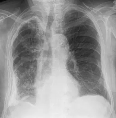 Case #12429420Chest · Intermediate
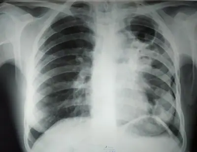 Case #76056976Chest · Intermediate
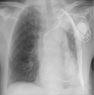 Case #110321807Chest · Intermediate
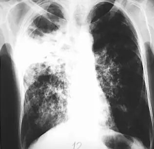 Case #5825373Chest · Intermediate
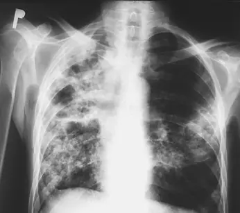 Case #5825382Chest · Intermediate
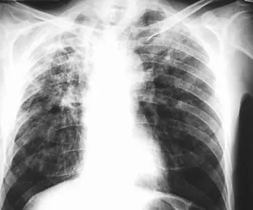 Case #5825390Chest · Intermediate
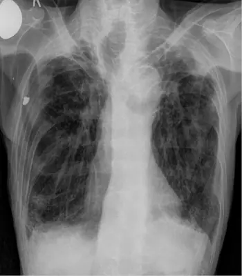 Case #107260282Chest · Intermediate
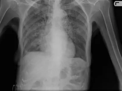 Case #31127199Chest · Intermediate
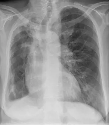 Case #111554897Chest · Intermediate
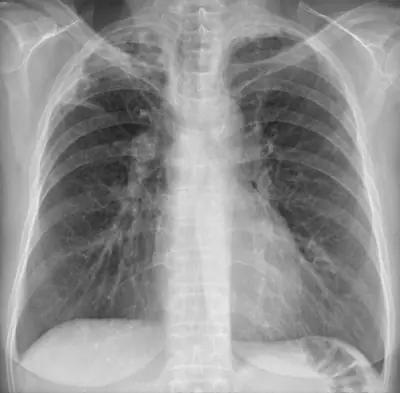 Case #144613371Chest · Intermediate
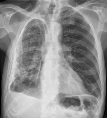 Case #36712504Chest · Intermediate
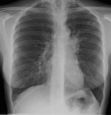 Case #3189243Chest · Intermediate
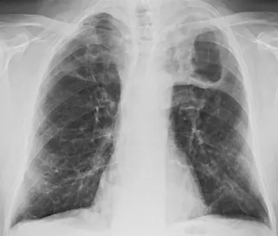 Case #95478070Chest · Intermediate
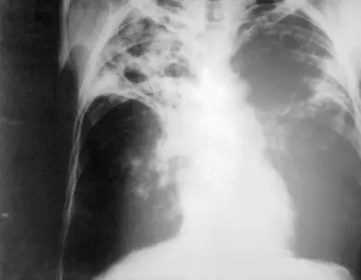 Case #880747Chest · Intermediate
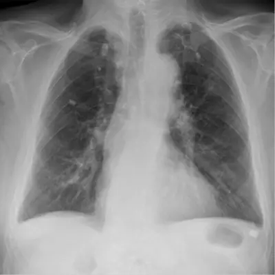 Case #107524233Chest · Intermediate
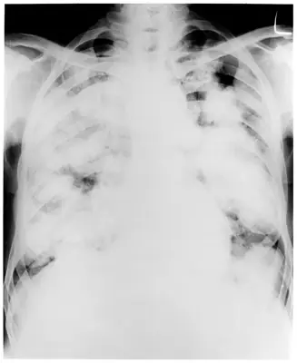 Case #36340193Chest · Intermediate
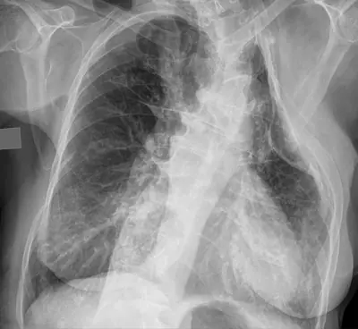 Case #139958909Chest · Intermediate
Other chest diagnoses
- Asbestos-related pleural disease
- COVID-19 pneumonia
- Cardiomegaly
- Cervical rib
- Diaphragmatic hernia
- Emphysema / COPD (hyperinflation)
- Hiatus hernia
- Lung abscess
- Lung cancer (mass)
- Lung collapse (atelectasis)
- Normal chest radiograph
- Pectus excavatum
- Pericardial effusion
- Pleural effusion
- Pneumocystis pneumonia (PCP)
- Pneumomediastinum
- Pneumonia (consolidation)
- Pneumothorax
- Pulmonary oedema (heart failure)
- Sarcoidosis
- Silicosis / pneumoconiosis
- Thoracic aortic aneurysm
Diagnoses come from each image's documented source. Credits and licences are shown on each case and on the image credits page.