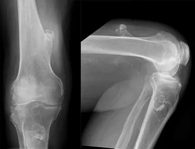
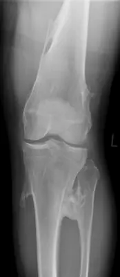
Case: Bone & joint disease radiograph #118625313
Bone & joint disease Beginner
Read this radiograph systematically before revealing the answer. What is the main abnormality?
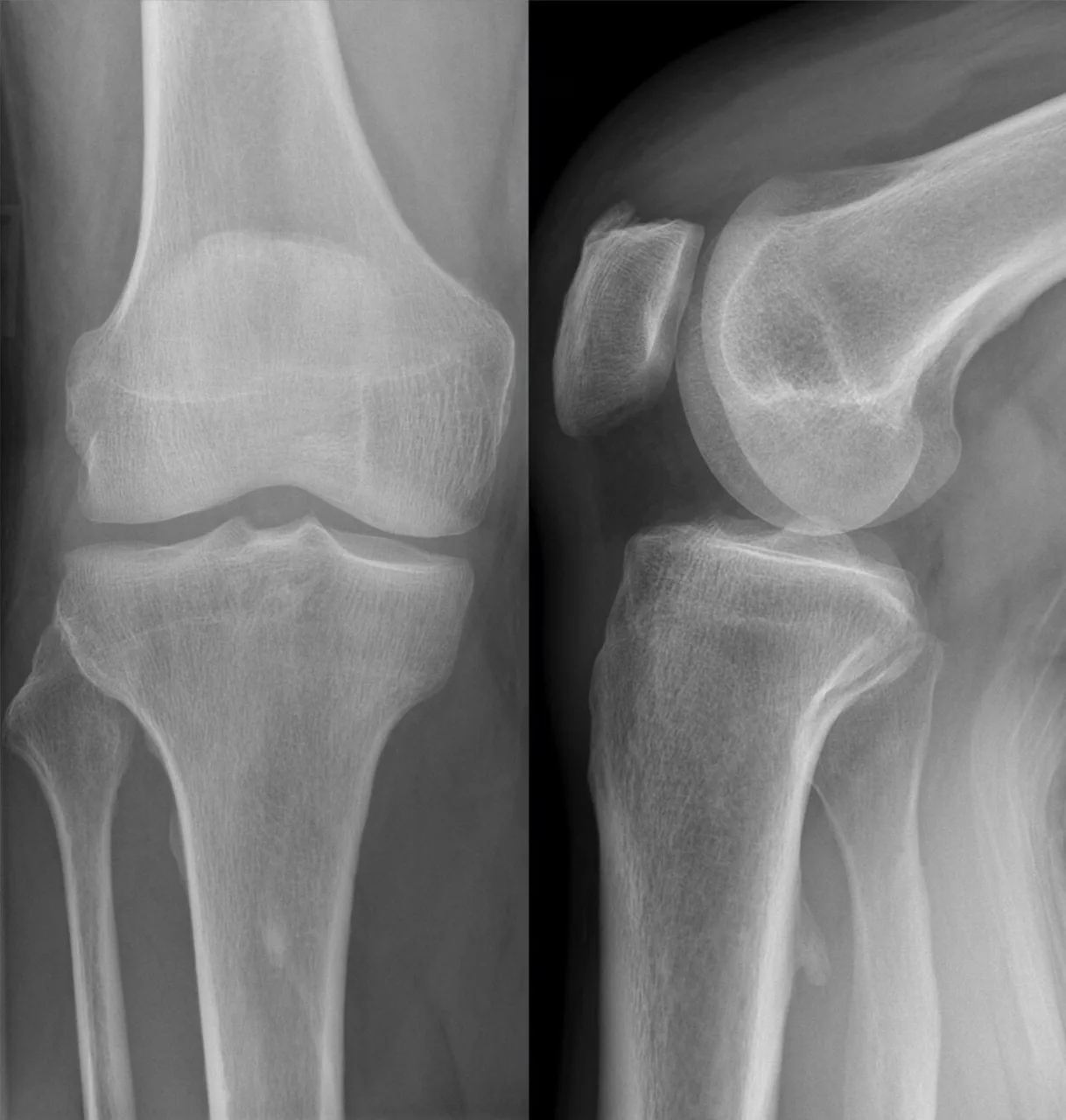
Image: Hellerhoff via Wikimedia Commons · CC BY-SA 4.0 · source · resized and converted to WebP
Tip: scroll or pinch to zoom, drag to pan, and try Invert.
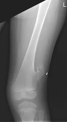
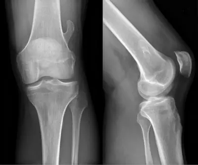
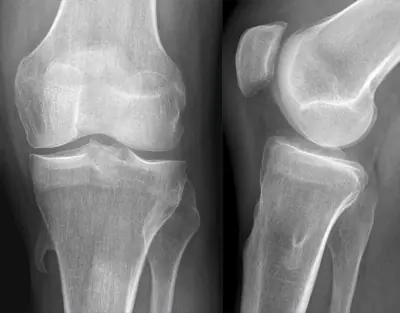
Answer: Osteochondroma (exostosis)
What to look for in osteochondroma (exostosis)
- Pedunculated exostosis with cortical continuity, pointing away from the joint.
Description by the image author (may be in another language): Exostose dorsal am proximalen Tibiaschaft, abgerundete hakenförmig nach distal weisend ähnlich wie ein Processus supracondylaris am Humerus. Insgesamt kein typisches Bild wie bei kartilaginärer Exostose.
Educational case. The diagnosis comes from the image's documented source; the "what to look for" list describes the condition in general and is not a report of this individual image.