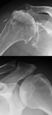
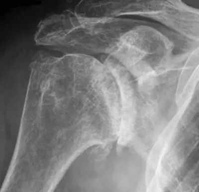
Case: Bone & joint disease radiograph #38256291
Bone & joint disease Beginner
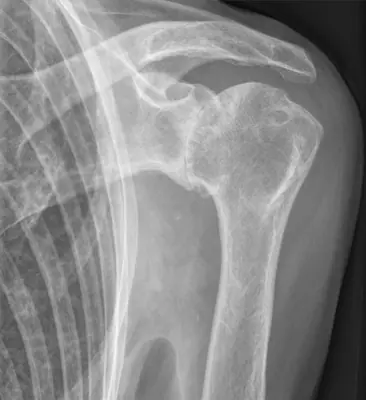
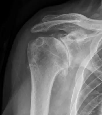
Read this radiograph systematically before revealing the answer. What is the main abnormality?
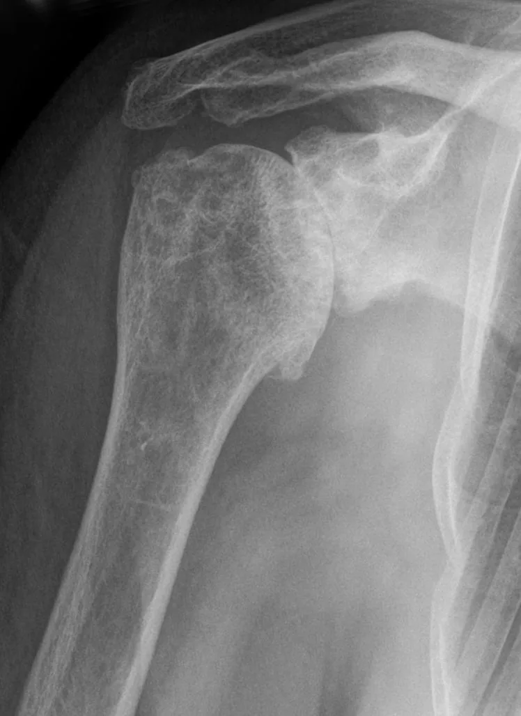
Image: Hellerhoff via Wikimedia Commons · CC BY-SA 3.0 · source · resized and converted to WebP
Tip: scroll or pinch to zoom, drag to pan, and try Invert.
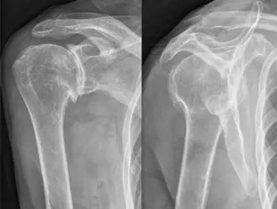
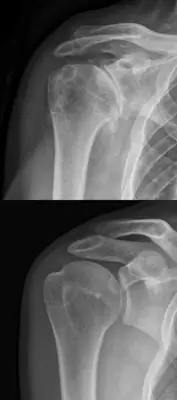
Answer: Osteoarthritis
What to look for in osteoarthritis
- Loss of joint space, typically uneven (non-uniform).
- Osteophytes at the joint margins.
- Subchondral sclerosis and subchondral cysts (the four signs: LOSS).
Description by the image author (may be in another language): Omarthrose, also Arthrose des Schultergelenks in der Röntgenaufnahme. Praktisch aufgehobener Gelenkspalt und vermehrte Sklerosierung der eigentlich knorpeltragenden Knochenoberflächen des Humerus und des Glenoids. Beginnende intraossäre Zystenbildung und ossäre Ausziehungen vor allem am Unterrand des Humerus.
Educational case. The diagnosis comes from the image's documented source; the "what to look for" list describes the condition in general and is not a report of this individual image.