Osteoporosis on X-ray: 3 real cases
Real, openly licensed radiographs showing osteoporosis. Study what to look for, then open each case and read it yourself before revealing the answer.
What to look for
- Generalised reduction in bone density with thin cortices.
- Prominent vertical trabeculae in vertebral bodies and fragility fractures (wedge fractures, hip, wrist).
- Plain film is insensitive: around 30% or more of bone mass may be lost before it is visible; DXA is the diagnostic test.
Quiz yourself on bone & joint disease images
Cases
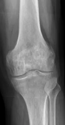 Case #97158442Bone & joint disease · Beginner
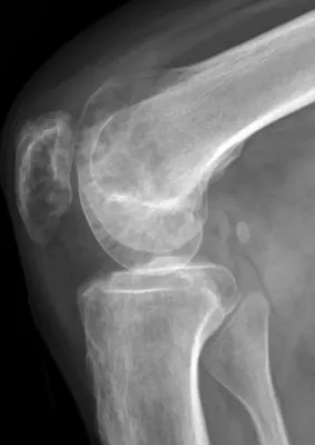 Case #97158668Bone & joint disease · Beginner
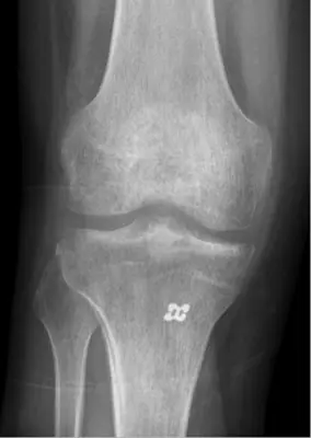 Case #96134114Bone & joint disease · Beginner
Other bone & joint disease diagnoses
- Avascular necrosis of the femoral head
- Bone metastases
- Enchondroma
- Freiberg disease
- Gout
- Hallux rigidus (first MTP osteoarthritis)
- Hallux valgus (bunion)
- Heterotopic ossification
- Knee osteoarthritis
- Multiple myeloma
- Neuropathic (Charcot) arthropathy
- Non-ossifying fibroma
- Osteoarthritis
- Osteochondroma (exostosis)
- Osteoma
- Osteomyelitis
- Rheumatoid arthritis
- Synovial chondromatosis
Diagnoses come from each image's documented source. Credits and licences are shown on each case and on the image credits page.