Femoral shaft fracture on X-ray: 8 real cases
Real, openly licensed radiographs showing femoral shaft fracture. Study what to look for, then open each case and read it yourself before revealing the answer.
What to look for
- A fracture through the femoral diaphysis, usually from high-energy trauma in adults.
- Describe pattern, displacement, angulation and shortening.
- Image the hip and knee too; an ipsilateral femoral neck fracture can be missed.
Quiz yourself on lower limb images
Cases
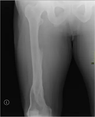 Case #17326603Lower limb · Beginner
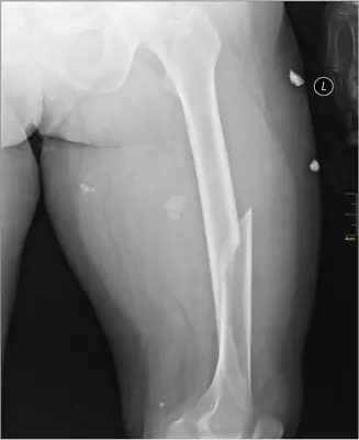 Case #17373724Lower limb · Beginner
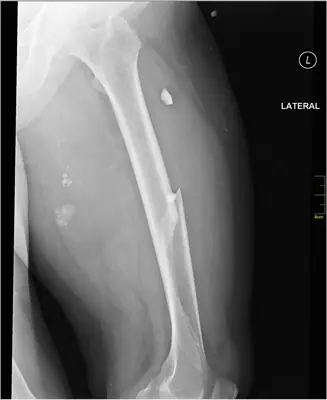 Case #17373764Lower limb · Beginner
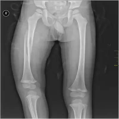 Case #17738088Lower limb · Beginner
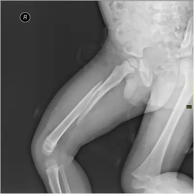 Case #17738090Lower limb · Beginner
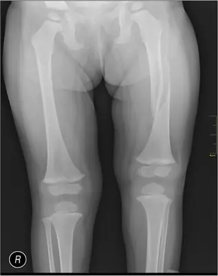 Case #17755157Lower limb · Beginner
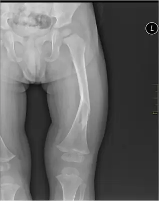 Case #17755369Lower limb · Beginner
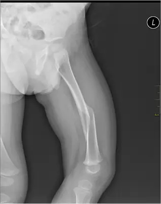 Case #17755371Lower limb · Beginner
Other lower limb diagnoses
- Acetabular fracture
- Ankle fracture (lateral malleolus)
- Bimalleolar ankle fracture
- Calcaneal fracture
- Calcaneal spur
- Fifth metatarsal fracture (Jones / base)
- Hip (proximal femoral) fracture
- Hip dislocation
- Knee joint effusion
- Patellar dislocation
- Patellar fracture
- Pellegrini–Stieda lesion
- Stress fracture
- Tibial (and fibular) shaft fracture
- Tibial plateau fracture
Diagnoses come from each image's documented source. Credits and licences are shown on each case and on the image credits page.