Calcific tendinitis on X-ray: 6 real cases
Real, openly licensed radiographs showing calcific tendinitis. Study what to look for, then open each case and read it yourself before revealing the answer.
What to look for
- Amorphous, cloud-like calcification in a tendon, most often supraspinatus just above the greater tuberosity.
- No fracture line and no bony cortex around the density.
- Calcifications can change size or disappear over time.
Learn: Shoulder and clavicle fractures
Quiz yourself on upper limb images
Cases
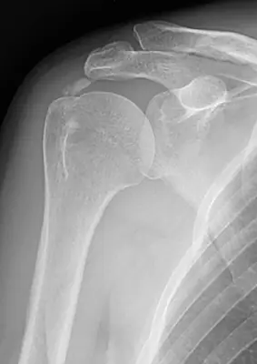 Case #4881675Upper limb · Intermediate
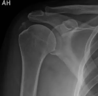 Case #49201531Upper limb · Intermediate
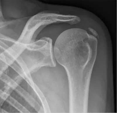 Case #110482682Upper limb · Intermediate
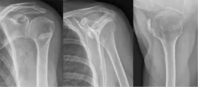 Case #105284026Upper limb · Intermediate
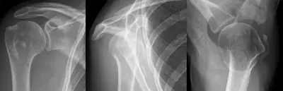 Case #119888244Upper limb · Intermediate
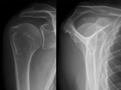 Case #146352243Upper limb · Intermediate
Other upper limb diagnoses
- Acromioclavicular joint dislocation
- Carpal or carpometacarpal dislocation
- Clavicle fracture
- Distal radius fracture
- Elbow dislocation
- Finger (interphalangeal) dislocation
- Finger (phalangeal) fracture
- Forearm shaft fracture (radius and ulna)
- Galeazzi fracture-dislocation
- Hill-Sachs lesion
- Humeral shaft fracture
- Metacarpal fracture
- Proximal humerus fracture
- Radial head fracture
- Rolando fracture
- Scaphoid fracture
- Shoulder (glenohumeral) dislocation
Diagnoses come from each image's documented source. Credits and licences are shown on each case and on the image credits page.