Case: Chest radiograph #162791180
Chest Advanced
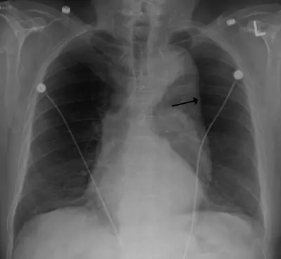
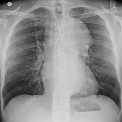
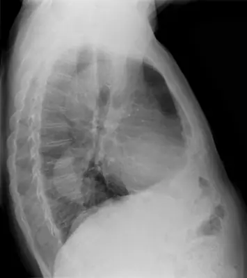
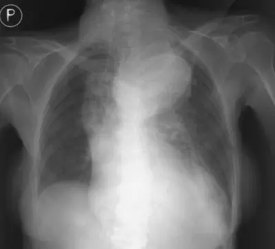
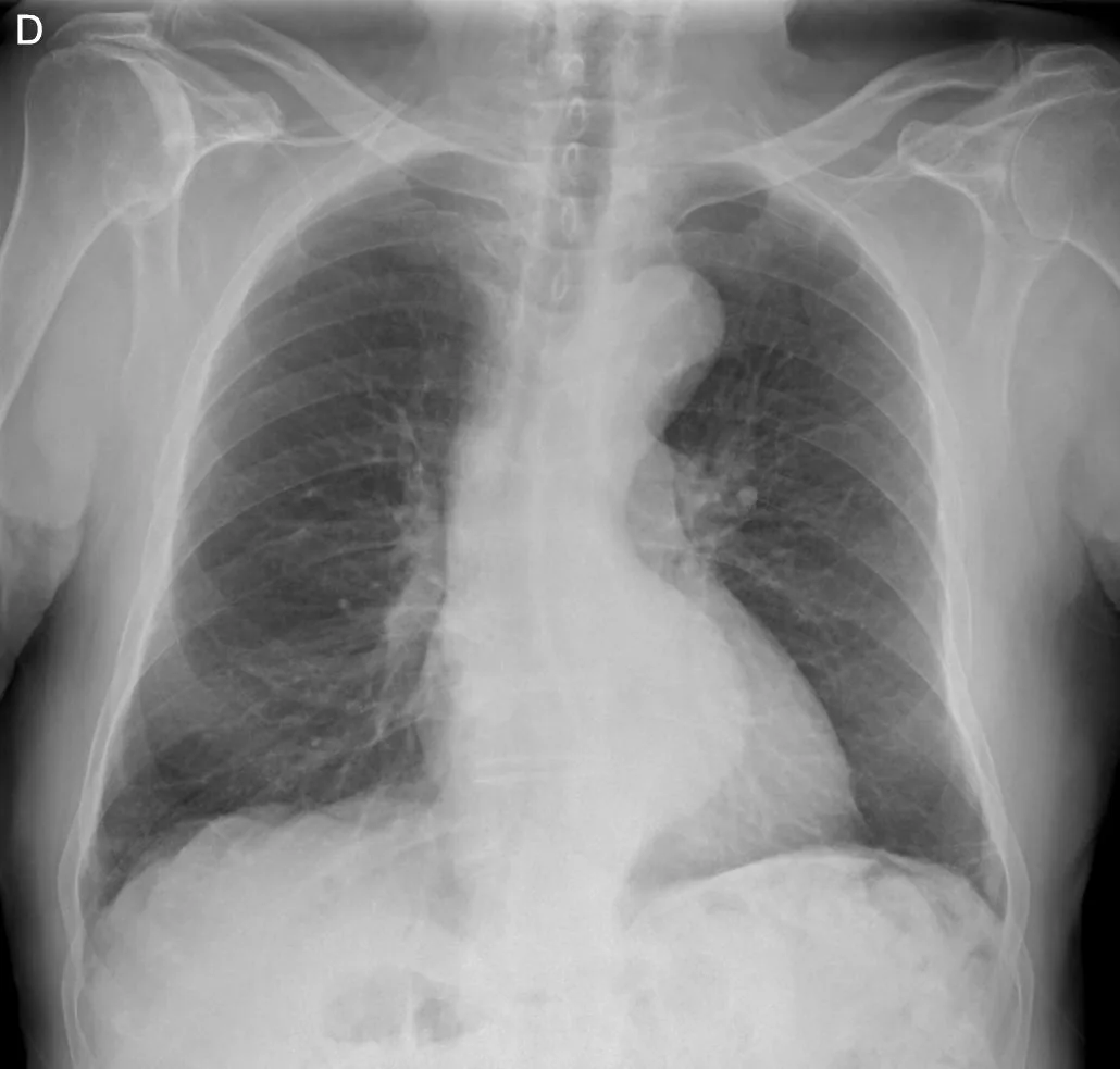
Read this radiograph systematically before revealing the answer. What is the main abnormality?
Tip: scroll or pinch to zoom, drag to pan, and try Invert.
Answer: Thoracic aortic aneurysm
What to look for in thoracic aortic aneurysm
- Widened or abnormally prominent aortic contour (knuckle or descending aorta).
- Mediastinal widening that follows the course of the aorta.
- Curvilinear wall calcification may outline the dilated aorta.
- Plain film cannot exclude or characterise aneurysm or dissection; CT angiography is required.
Description by the image author (may be in another language): Posteroinferior thoracic aortic aneurysm, frontal X ray. 91-years-old man.
Educational case. The diagnosis comes from the image's documented source; the "what to look for" list describes the condition in general and is not a report of this individual image.